![]()

High Resolution Ultrasound Imaging and Doppler Assessment in Mice
|
||
|
We use the micro-ultrasound system developed by Dr. Stuart Foster and commercialized by VisualSonics. The ultrasound biomicroscope generates high resolution real-time images of living embryos in utero, and detects strong blood flow (Doppler) signals even from tiny embryonic mouse hearts <1 mm in size. We do echocardiographic exams similar to those performed clinically on embryos, neonates and adult mice. We also use micro-ultrasound to guide fine-tipped cannulae into precise locations within the developing conceptus as young as 6.5 d gestation (2 days postimplantation), and to inject nl volumes into targets only 200 μm in size. We are using these methods to develop treatments to promote optimal growth and development of the fetus and placenta. In complicated human pregnancies, we hope these treatments will one day lead to the birth of healthier babies with brighter futures.
|
||
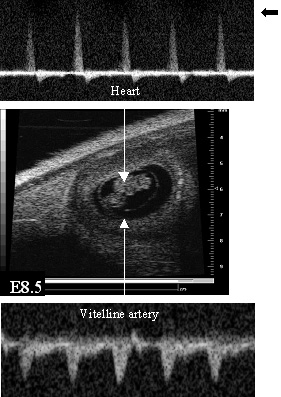 |
Doppler blood velocity in the heart (top) and in the vitelline artery to the yolk sac of a mouse embryo at 8.5 days of gestation (bottom) on the day the primordial heart starts to beat. The placenta has not developed yet. The image in the middle shows the embryo inside the uterus. By Dr. Junwu Mu in the Adamson Lab.
|
|


.")

